 GOUT
is caused by monosodium urate or uric acid crystal
deposition within cartilage, bone, or periarticular tissues.
GOUT
is caused by monosodium urate or uric acid crystal
deposition within cartilage, bone, or periarticular tissues.
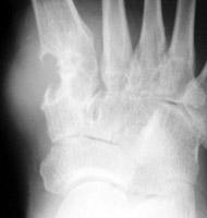
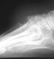
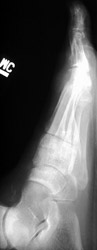
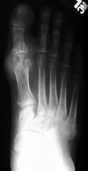
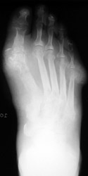
1. Distribution
First metatarsophalangeal joint is most commonly affected, followed by the
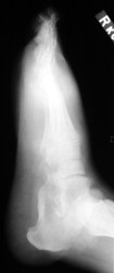
first interphalangeal joint and tarsometatarsal joints. Posterior calcaneal
involvement has also been noted. The majority of first presentations are monoarticular.
Bilateral and symmetric or asymmetric polyarticular involvement may be present
within any of the foot joints.
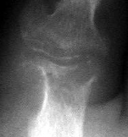
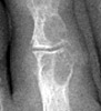
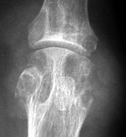
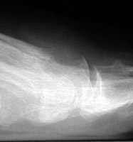
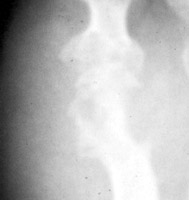
2. Erosion pattern:
Acute, episodic soft tissue swelling may represent the earliest radiographic
sign. Later, sharp, round or oval marginal joint erosions with sclerotic borders
are classically seen with gout. These findings most commonly occur along the
dorsum of the foot. Associated soft tissue tophi or intraosseous nodules may
be present. "Overhanging margin" occur where the bone resorbs beneath a tophaceous
nodule. Joint spaces are usually preserved, but ankylosis may rarely occur
with advanced stages of gout. The aforementioned findings may be in different
stages of progression with any given patient.
3. Differential diagnosis:
Concomitant osteoarthritis (especially of the tarsometatarsal joint) is common
and should not be confused with gout. Joint space narrowing, as seen with
advanced gout, may mimic rheumatoid arthritis.