![]()
![]()
![]()
![]()
![]()
![]()
![]()
![]()
![]()
![]()
![]()
![]()
![]()
|
|
|
t
Pigmented Villonodular SynovitisIn this entity, there is synovial proliferation and hemosiderine deposition.
Divided into localized or nodular types. Mass usually arise in the synovial
lining of the joints, tendon sheaths, fascial planes, or ligamentous tissues. It
is usually monoarticular and most commonly seen in young adult men. Hand and
feet are frequently involved in localized form, and knee is the common location
in the diffuse pigmented villonodular synovitis.
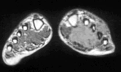
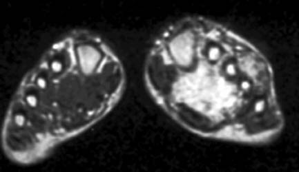
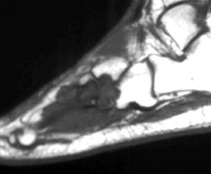
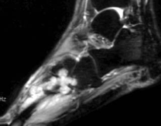
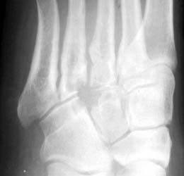
26 years old female presenting with painful soft tissue mass at the plantar aspect of the left foot.MR demonstrates this mass is isointense on T1,isointense to minimally hyperintense on proton density and hyperintense on T2 W and Stir images. Bony erosions are identified in 3rd cuneiform, cuboid and bases of 3rd and 4th metatarsals on MRI and plain radiographs.Few foci of low signal intensity hemosiderine deposits are identified on all pulse sequences. |
|
|